
A NEW INSTRUMENT WILL INSPECT WHOLE EYE WITH GOOD INTERPRETATION
Dr,DRAM,HIV /AIDS,HEPATITIS ,SEX DISEASES & WEAKNESS expert,New Delhi,India,+917838059592
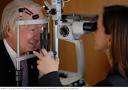
Researchers have developed the first instrument that can provide a detailed image of the entire eye. By incorporating a lens that changes optical parameters in response to an electric current, the innovative technology can produce higher quality images than currently available and could make eye examinations faster and more comfortable for patients by avoiding the need to undergo imaging with multiple instruments to look at different areas of the eye.
“An instrument that can examine the whole eye will improve the patient’s experience because they won’t have to go through imaging with different devices. It might also one day reduce the number of instruments — which can be quite expensive — needed in an ophthalmology clinic.”In Optica, The Optical Society's journal for high impact research, the researchers show that their new optical coherence tomography (OCT) imaging system can not only image both the front and the back of the eye, but can also image the interfaces of the eye’s vitreous gel with the retina and lens with unprecedented detail. This new imaging capability could allow scientists to better understand how the vitreous gel that fills the eye interacts with the retina and why it can sometimes become detached with aging.
Researchers developed a new OCT system that can image the entire eye. “We also want to use our instrument to measure opacities in the eye’s crystal lens and the vitreous to better understand how various parts of the eye affect the deterioration of vision,” said Grulkowski. “We believe that the ability to measure these opacities and other properties of the eye that couldn’t be examined before will open up many new ophthalmology applications for OCT.”
Increasing imaging depth
The new system is based on OCT, which is commonly used to acquire very detailed, cross-sectional ophthalmology images. Most clinical instruments are limited to imaging depths of 2 to 3 millimeters, and it is difficult to switch between imaging the front and back portions of the eye because the eye is composed of elements that bend the light to focus it onto the retina.To overcome these challenges, the researchers used an electrically tunable lens to build an OCT instrument that could focus light in a way that enabled whole-eye imaging. Unlike standard glass or plastic lenses, which have fixed parameters, the optical properties of an electrically tunable lens can be dynamically controlled using an electric current.
The researchers used their new system to measure the anatomical characteristics of the eyes of seven healthy people. Measurements calculated using images from the new system correlated well with those obtained with an ocular biometer, the standard clinical device used today.
The researchers are now working to optimize the instrument for imaging of the entire vitreous gel, not just where it interfaces with the lens and retina. The vitreous gel has not been studied intensively and is difficult to image because it is highly transparent. The ability to image the entire vitreous could allow OCT to be used to guide procedures that involve the removal of the vitreous gel from the eye, which is sometimes done to repair retinal detachment.
 Comments (
 Category (
 Views (
- Kidney stones universally present hazard in north india,dillution by water prevent it
- Steroid and placebo effect equally for mild persisting asthma with low sputum eosinophils
- Government wants to fix public healthcare staff shortages with ayush docs: will it work?
- Plea in hc for payment of salaries of edmc, north mcd teachers and doctors
- 7 indian pharma companies named in us lawsuit over inflating generic drug prices
- Woman in up dies after explosion in her mouth during treatment,what is diagnosis?
- Woman in up dies after explosion in her mouth during treatment,what is diagnosis?
- Woman in up dies after explosion in her mouth during treatment,what is diagnosis?
- Air pollution ! mothers organising rally in london,anaesthetist choosing gas,will india follow?
- Cardiac arrest is always not sudden as understood -a study
News & Highlights | Articles | Drug Magic | Community Weblog | Health Manager | Testimonials | Video Gallery | Medical Humour
Home | About 'India HeartBeat' | Awards | Contact Us | Terms of Use | Privacy Policy | Disclaimer | Advertise With Us | FAQs | Links | Quick Tour